Secret biology of corals unveiled by groundbreaking microscope
Groundbreaking new tech has made it possible to study the health and physiology of coral reefs in their natural habitat, advancing efforts to better understand processes such as coral bleaching currently affecting swathes of ocean.

The intricate, hidden processes that sustain coral life are being revealed through a new microscope developed by scientists at UC San Diego’s Scripps Institution of Oceanography, advancing efforts to understand precisely why corals bleach.
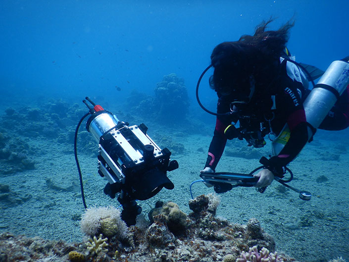
The diver-operated microscope – called the Benthic Underwater Microscope imaging PAM (or BUMP) – uses something called ‘pulse amplitude modulated (PAM) light techniques to “offer an unprecedented look at coral photosynthesis” on a microscopic level.
The technology makes it possible for researchers to study the health and physiology of coral reefs in their natural habitat, advancing those longstanding efforts to better understand processes such as coral bleaching currently sweeping across swathes of reef ecosystems.
The cutting-edge microscope has been built – with funding from the US National Science Foundation – by a team of engineers and marine researchers in the Jaffe Lab for Underwater Imaging at Scripps Oceanography. Already, the microscope is yielding new insights into the relationship between corals and the symbiotic micro-algae that support their health.
Such level of microscopic research has revealed – for the first time – just how well individual algae photosynthesise within its host’s coral tissue.
The findings have been published this week in the journal Methods in Ecology and Evolution.
“This microscope is a huge technological leap in the field of coral health assessment,” said Or Ben-Zvi, a postdoctoral researcher at Scripps Oceanography and lead author of the study. “Coral reefs are rapidly declining, losing their photosynthetic symbiotic algae in the process known as coral bleaching.
“We now have a tool that allows us to examine these microalgae within coral tissue, non-invasively, and in their natural environment.”
In other news


Corals are reef-building animals that can’t photosynthesise on their own. Instead, they rely on micro-algae living inside their tissues to do it for them. These symbiotic algae use sunlight, carbon dioxide, and water to produce oxygen and energy-rich sugars that support coral grwoth and reef formation.
At around 10 micro-metres across – about one-tenth the width of a human hair – these algae are far too small to be seen with the naked eye. When corals are stressed by warming waters or poor environmental conditions, they lose these micro-algae, leading to a pale appearance.
This is the process known as coral bleaching. It leads – eventually – to the starvation of the coral. Although this process is known, scientists don’t fully understand why, and it hasn’t been possible to study the process at appropriate scales in the field – until now.
“The microscope facilitates previously unavailable, underwater observations of coral health, a breakthrough made possible thanks to the National Science Foundation and its critical investment in technology development,” said Jules Jaffe, a research oceanographer at Scripps and co-author of the study.
“Without continued federal funding, scientific research is jeopardised. In this case, NSF funding allowed us to fabricate a device so we can solve the physiological mystery of why corals bleach, and ultimately, use these insights to inform remediation efforts.”
Through an array of high-magnification lenses and focused LED lights, the microscope captures vivid colour and fluorescence images and videos. It also now has the ability to measure photosynthesis and map it in higher resolution via focal scans. Scientists can use this to create high-resolution 3D scans of corals.
Working in collaboration with the Smith Lab at Scripps Oceanography, Ben-Zvi – a marine biologist – has tested and calibrated the instrument at several coral reef hotspots around the globe, including in Hawaii, the Red Sea, and Palmyra Atoll.
Throughout her many observations, Ben-Zvis has been most surprised by how active the corals have been, noting that they changed their volume and shape constantly. She even observed instances in which a coral polyp appeared to be trying to capture or remove a particle that was passing by, by rapidly contracting its tentacles.
“The more time we spend with this microscope, the more we hope to learn about corals and why they do what they do under certain conditions,” said Ben-Zvi. “We are visualising photosynthesis, something that was previously unseen at the scales we are examining, and that feels like magic.”
The non-invasive technique allows researchers to assess the health of corals without the need to interrupt nature – it’s similar, Ben-Zvi has said “to checking on the coral’s pulse without giving them a shot or doing an intrusive procedure on them.”
The researchers have also said that data collected with the new microscope can reveal early warning signs that appear before corals experience irreversible damage from global climate change events, such as marine heat waves. These insights could help guide mitigation strategies to better protect them.
Beyond corals, and the tool has other widespread potential for studying other small-scale marine organisms that photosynthesise, such as baby kelp. In fact, several researchers at Scripps Oceanography are already using the BUMP imaging system to study the early life stages of the exclusive giant kelp off California.
“Since photosynthesis in the ocean is important for life on earth, a host of other applications are imaginable with this tool, including right here off the coast of San Diego,” said Jaffe.


"*" indicates required fields
Printed editions
Current issue
Back issues


Back Issues
Issue 43 Sir David Attenborough’s ‘Ocean’


Back Issues
Issue 41 Holdfast to the canopy
Enjoy so much more from Oceanographic Magazine by becoming a subscriber.
A range of subscription options are available.
